Hair loss
Alopecia areata
An autoimmune condition in which the immune system attacks hair follicles, causing smooth round patches of hair loss on the scalp or beard, and in more extensive forms loss of all scalp hair or all body hair. It can affect children and adults, often starts suddenly and can regrow on its own, particularly in limited patches. Treatments include intralesional and potent topical corticosteroids, topical immunotherapy and, for extensive disease, oral small molecule inhibitors that block the immune signalling involved.
Androgenetic alopecia
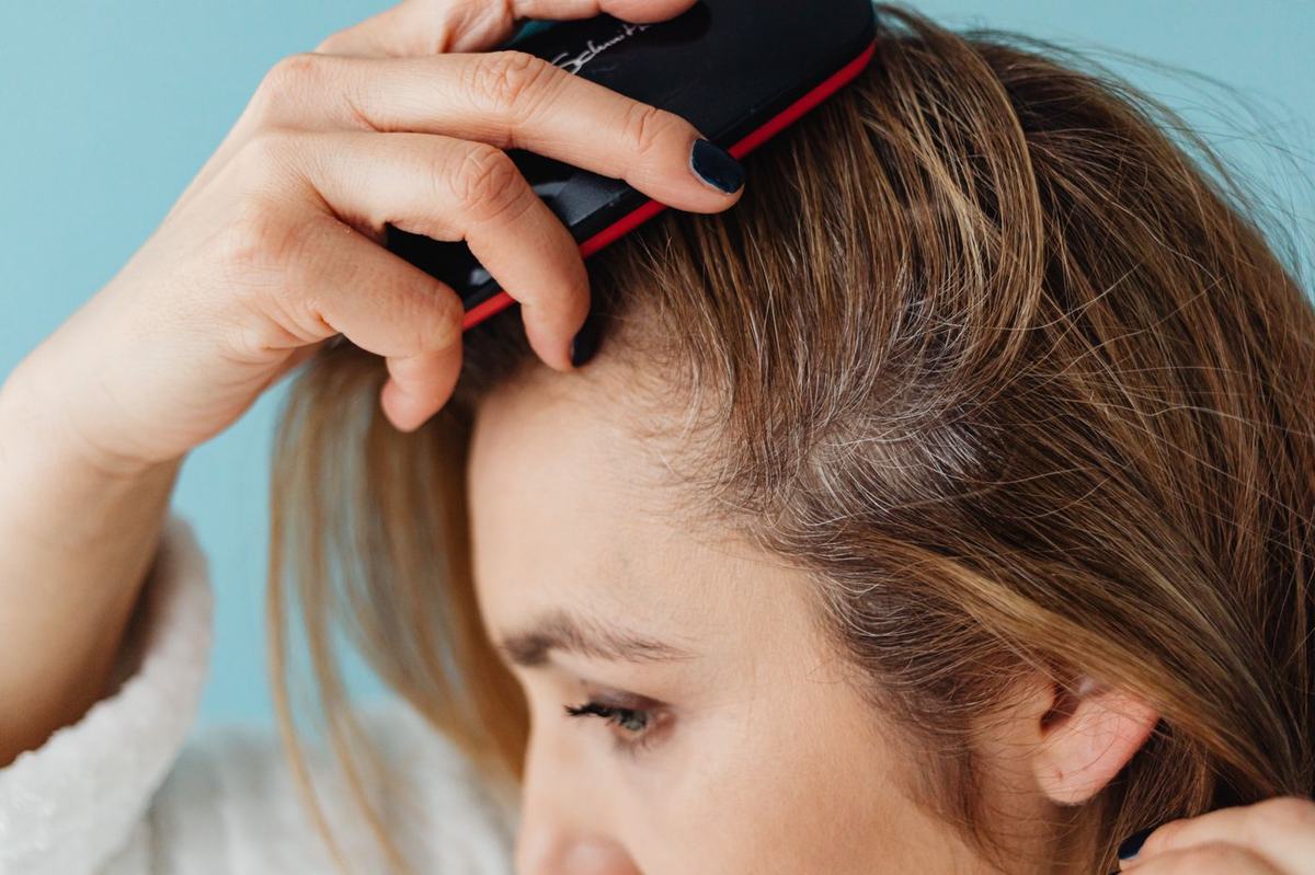
Pattern hair loss is the most common cause of thinning in both men and women. It is genetic and hormonally driven, and gradual. In men it recedes at the temples and crown; in women it thins across the top of the scalp with the front hairline usually kept. Topical and oral treatments can slow loss and partially regrow hair, and must be continued to maintain the effect. In women, tests may be done to rule out iron deficiency, thyroid disease or hormonal disorders that can contribute.
Scarring alopecias
In this group inflammation destroys the follicle and replaces it with scar, so hair lost is permanent. Examples include lichen planopilaris, frontal fibrosing alopecia, discoid lupus of the scalp and central centrifugal cicatricial alopecia. Redness, scale, itch or tenderness around the hair roots, or a shiny, smooth appearance of the bald area, are clues. Because the aim is to halt progression before more follicles are lost, prompt referral and a scalp biopsy to confirm the diagnosis are important. Treatment is with anti-inflammatory therapy, topical, injected or oral, depending on the type and activity.
Diffuse shedding
Widespread shedding two to four months after an illness, surgery, childbirth, rapid weight loss or a new medication is usually telogen effluvium, which settles once the trigger has passed. Assessment confirms the cause and rules out other conditions.
Scalp conditions
Scalp psoriasis produces thick, well defined scaly plaques that often extend past the hairline. Seborrhoeic dermatitis causes greasy, yellowish scale with redness in the scalp, eyebrows, beard and creases of the face, and is the cause of most dandruff. The two can look similar and sometimes overlap. Treatment includes medicated shampoos, scalp lotions or foams containing corticosteroids or vitamin D analogues, antifungal preparations, and preparations to lift heavy scale. See also psoriasis.
Nail conditions
Nail psoriasis
Pitting, yellow brown discolouration, thickening, crumbling and lifting of the nail from its bed are typical. Nail involvement is associated with a higher likelihood of psoriatic arthritis, so joint symptoms are asked about. Treatment can be slow because nails grow slowly, and options include potent topical therapy, corticosteroid injection at the nail fold and, where skin or joints are also affected, systemic therapy.
Fungal nail infection
Onychomycosis causes thickened, discoloured, crumbly nails, most often the toenails. Because several conditions mimic it, the diagnosis is confirmed with nail clippings sent for microscopy and culture before oral antifungal treatment is prescribed. Oral therapy for several months is the most effective option; topical lacquers suit milder cases. Recurrence is common and preventive measures are discussed.
Nail changes as a sign of disease
Nails can reflect what is happening elsewhere. A dark streak running the length of a nail, particularly a single nail in an adult, or pigment spreading onto the surrounding skin, needs assessment to exclude melanoma of the nail unit. Clubbing, spoon shaped nails, horizontal ridges and splinter haemorrhages may point to lung, heart, blood or connective tissue disease, and your dermatologist will arrange or recommend further investigation where indicated.
Assessment at DermCare
Assessment starts with a history of onset, pattern, family history, general health, medications and hair or nail care practices, followed by examination of the scalp, hair, nails and skin. Dermoscopy of the scalp and nails adds detail. Blood tests are arranged where a contributing condition is suspected. Where the diagnosis remains uncertain, particularly in suspected scarring alopecia or an unusual nail change, a small biopsy is taken under local anaesthetic and read in house by a dermatologist who is also certified in Anatomical Pathology.
What to bring
Images of your scalp or nails from earlier months or years help show the pace of change. Bring a list of medications and supplements, and the names of any shampoos, lotions or nail treatments you have already tried.
For referring physicians
Please describe the pattern and duration of loss, any symptoms of scalp inflammation, family history and relevant blood work already done, including ferritin and thyroid function where obtained. Suspected scarring alopecia and any pigmented nail streak should be flagged for earlier assessment. For nail dystrophy, please state whether mycology has been sent. How to refer.