Types of skin cancer
Basal cell carcinoma is the most common type. It usually appears on sun exposed skin as a pearly or translucent bump, a flat pink patch, or a sore that bleeds and does not heal. It rarely spreads but can grow into surrounding tissue if left.
Squamous cell carcinoma often appears as a firm, scaly or crusted lump, sometimes tender, on the face, scalp, ears, lips or backs of the hands. It carries a small risk of spread, higher in people who are immunosuppressed.
Melanoma arises from pigment cells and can develop in an existing mole or as a new dark spot. It is less common than the other types but more serious, because it can spread if not removed early. Melanoma can also appear in people with darker skin, often on the palms, soles or under the nails.
Actinic keratoses are rough, scaly patches caused by cumulative sun damage. They are not cancer, but a small proportion progress to squamous cell carcinoma, so they are treated and monitored.
Warning signs
For moles and pigmented spots, the ABCDE checklist is a useful guide.
- Asymmetry: one half does not match the other
- Border: edges are irregular, ragged or blurred
- Colour: more than one colour, or uneven shades of brown, black, pink or white
- Diameter: larger than about 6 mm, though melanomas can be smaller
- Evolving: any change in size, shape, colour or symptoms such as itch or bleeding
A spot that looks different from all your other moles, sometimes called the ugly duckling, deserves attention even if it does not meet the checklist. So does any sore that has not healed within a few weeks.
Assessment at DermCare
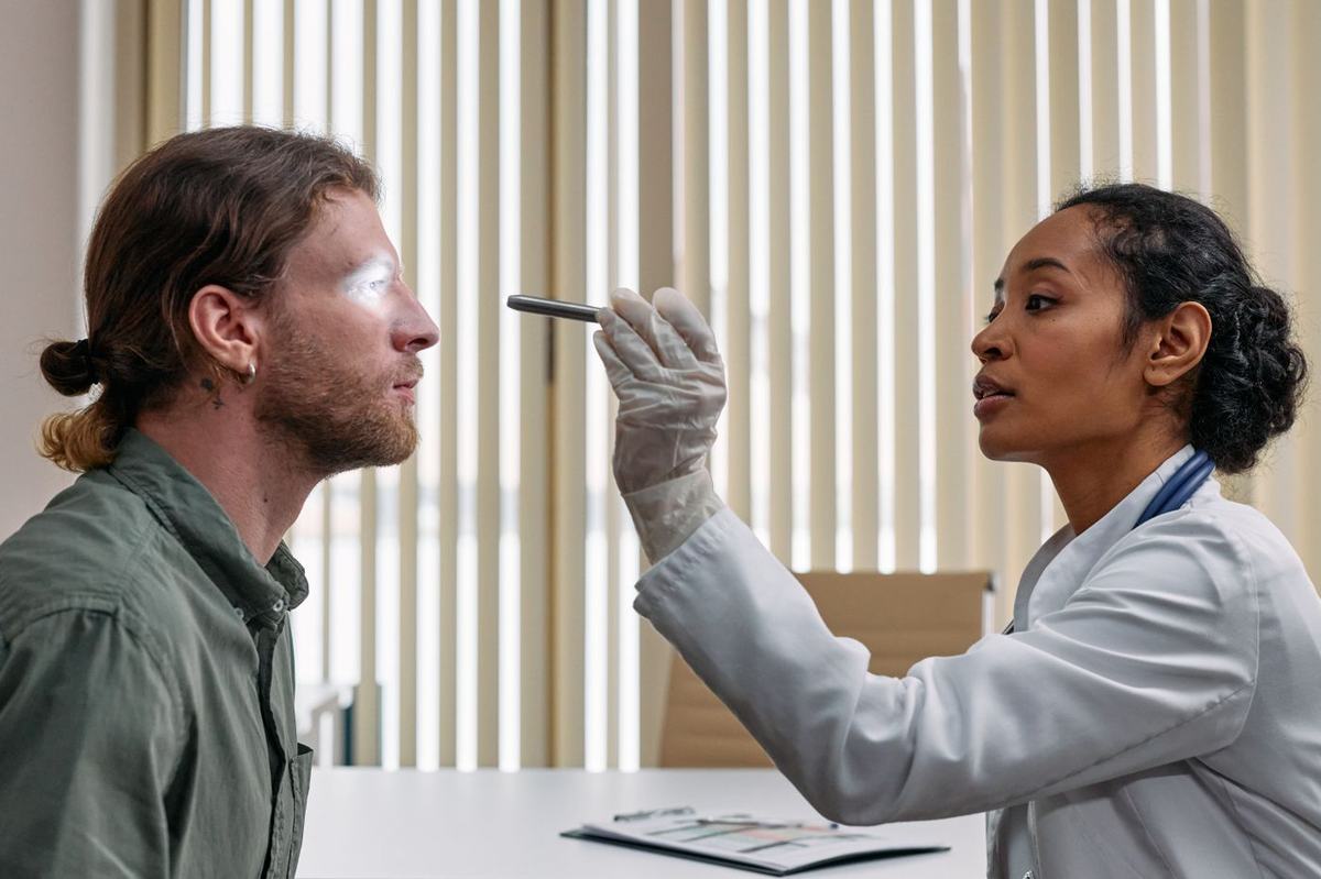
Your dermatologist examines the lesion of concern and, where appropriate, the rest of the skin. Dermoscopy, a handheld magnifier with polarised light, lets the dermatologist see structures below the surface that are not visible to the naked eye and improves the accuracy of diagnosis.
Where a lesion cannot be confidently called benign, a biopsy is taken. This is a short procedure under local anaesthetic in which all or part of the lesion is removed and sent for microscopic examination. Whether total body imaging or mole mapping is offered is being confirmed.
Dermatopathology in house
Biopsy specimens from DermCare are read in house by Dr. Faisal Al-Mohammedi, who is certified by the Royal College in both Dermatology and Anatomical Pathology. Having the clinician who saw the lesion and the pathologist who reads the slide in the same practice supports accurate correlation between what was seen on the skin and what is found under the microscope. Typical turnaround from biopsy to result is being confirmed.
Treatment options
Treatment depends on the type of lesion, its size and depth, where it is on the body, and your general health. Your dermatologist will explain the options and the reasons for the recommendation.
Surgical excision
Many skin cancers are treated by excision under local anaesthetic, removing the lesion with a margin of normal skin, which is then examined to confirm the cancer has been fully removed. The range of procedures performed on site at DermCare is being confirmed.
Other modalities
Superficial lesions and actinic keratoses may be treated with destructive methods such as cryotherapy or curettage, or with prescription topical therapy applied over a course of weeks. Field treatment is used where sun damage covers a wider area rather than a single spot.
Referral onward
Where a tumour is large, in a cosmetically or functionally sensitive site such as the eyelid or nose, or of a type that needs specialised surgery or oncology input, we refer onward to the appropriate surgical or cancer centre and continue to coordinate your skin care.
Sun protection
Once you have had one skin cancer or actinic keratoses, your risk of another is higher. Use a broad spectrum sunscreen with SPF 30 or above daily, wear a hat and protective clothing, avoid the midday sun and never use tanning beds. Check your own skin regularly and report changes.
For referring physicians
Please mark suspected melanoma, rapidly growing lesions and non healing ulcers as urgent and state why. Include the site, size, duration and any change, an image where possible, prior skin cancer history and immunosuppression. See the urgent triage criteria and how to refer.